Attribution 4.0 International (CC BY 4.0)
You can use this work for any purpose, including commercial uses, without restriction under copyright law. You should also provide attribution to the original work, source and licence. Read more about this licence.
Credit
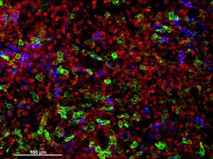
Cellular architecture of human skin lymphoma imaged by whole mount tissue microscopy. Normal human skin has a rich network of white blood cells (specifically dendritic cells, T cells and macrophages) which form sheaths around blood vessels. In diseased skin, such as in skin lymphoma as seen here, this normal architecture becomes distorted. In this image, lots of T cells (stained for CD3; red), dendritic cells (stained for CD11c; green) and macrophages (stained for LYVE-1; blue) have infiltrated the skin. X20 magnification. Scale bar (white) represents 100 micrometres. Dr. Xiao-nong Wang, Human Dendritic Cell Laboratory, Newcastle University. Attribution 4.0 International (CC BY 4.0). Source: Wellcome Collection.